The retina is an integral part of how the eyes work and function. If the retina isn’t working as it should, it can lead to severe issues with vision. At Coastal Surgical Center, our team aims to provide you with specialized care using the most state-of-the-art technology available.
What is the Retina?
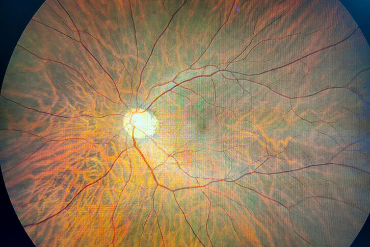
The retina is located at the back of the eye and is composed of several layers. In one of these layers, you’ll find photoreceptors.
There are two kinds of photoreceptors in the eye called rods and cones. Rods detect motion, allow you to see in black and white vision, and see well in low lighting. Cones enable you to have central and color vision and perform best in bright and medium lighting.
The photoreceptors in the retina take the light that the cornea and lens focus and turn them into signals that the brain then converts into the images that we see. If anything in the retina doesn’t work as it should, it leads to problems with your vision.
Retinal Detachment
One of the more common issues that the retina can face is something called retinal detachment. With a retinal detachment, a rip or tear forms in the retina.
A rip or tear often occurs because of pulling on the retina’s surface. Retinal detachments become more likely due to aging as the vitreous loses its flexibility and becomes more liquid.
Sometimes, when the vitreous pulls away from the retina, it can pull the torn retina away from the back of the eye. If left untreated, a retinal detachment can cause permanent vision loss.
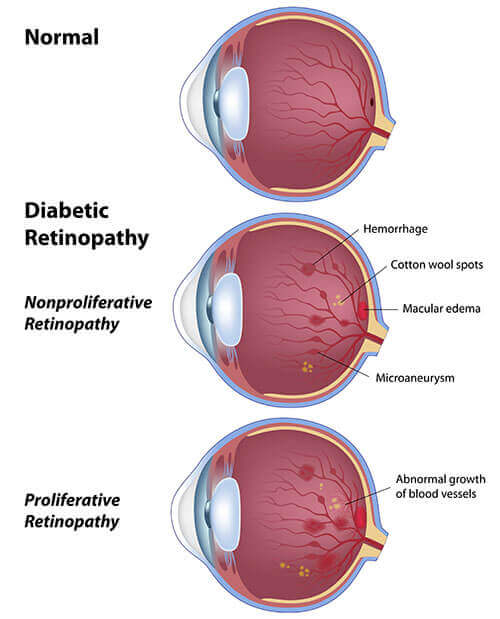
Diabetic Retinopathy
Patients with diabetes become more likely to develop diabetic retinopathy. Diabetic retinopathy occurs when high blood sugar levels damage blood vessels in the retina.
This damage can cause the vessels to leak and swell or close up, making it impossible for blood to pass through. There are two stages: non-proliferative diabetic retinopathy (NPDR) and proliferative diabetic retinopathy (PDR).
Non-proliferative diabetic retinopathy is the early stage of diabetic eye disease. With NPDR, blood vessels can close off in the retina, making blood unable to reach the macula. Particles called exudates may form on the retina and affect the vision.
With proliferative diabetic retinopathy, this is the more advanced stage of diabetic eye disease. It occurs when the retina begins growing newer, weaker blood vessels.
These blood vessels may bleed into the vitreous, causing a block of all vision. The new blood vessels can also form scar tissue and may eventually lead to a detached retina. PDR can also mean the loss of both your central and peripheral vision.
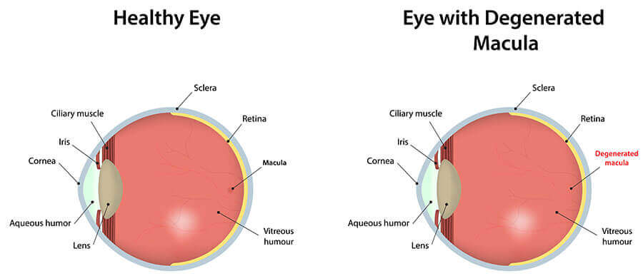
Macular Degeneration
Macular degeneration, also known as age-related macular degeneration (AMD), is the leading cause of vision loss in people 50 or older. AMD occurs when the macula becomes damaged, causing the loss of central vision.
There are two kinds of macular degeneration: dry and wet. Dry AMD is the most common kind since it affects almost 80% of patients with macular degeneration.
With dry AMD, the macula begins getting thinner because of age. As the macula thins, it causes small amounts of a protein called drusen to grow. As dry macular degeneration gets worse, you’ll slowly lose your central vision. Though there is no way to treat or cure dry AMD, some people may benefit from taking a specific combination of nutritional supplements.
Wet AMD is much rarer, but it happens when new abnormal blood vessels begin to grow under the retina. These new vessels may start leaking blood and other fluids.
When the blood vessels start leaking, it scars the macula and results in much faster vision loss than dry AMD. For patients with wet AMD, anti-VEGF medications can help reduce abnormal blood vessels in the retina while slowing down leaking.
Flashes and Floaters
Most of the time, seeing flashes and floaters isn’t worth worrying over. Floaters are parts of the vitreous that have solidified, causing particles called floaters to move and pass in front of the macula.
With age, the vitreous starts shrinking and creates these particles. Floaters can look like small shapes or small bits of dust that move with you.
Flashes are similar, but they usually look more like camera flashes or lightning. Flashes and floaters only become something to be concerned about if you start seeing a lot more flashes and floaters or you start seeing sudden flashes across your field of vision. If this occurs, it can be a sign of a retinal tear or a retinal detachment.
Are you looking to treat a retinal condition? Schedule an appointment at Coastal Surgical Center in Newington, NH, now!